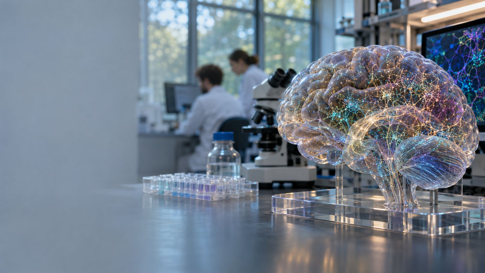
Basal Ganglia Atlas Offers the Most Detailed Map Yet
Basal Ganglia Atlas Offers the Most Detailed Map Yet
A New Map of a Disease-Linked Brain Region
Scientists have created a large-scale cellular atlas of the basal ganglia, a network of interconnected structures deep within the brain. The region is best known for regulating movement, but it also contributes to learning, motivation, reward processing, decision-making, and behavior. Disruptions in basal ganglia circuits are associated with neurological and psychiatric disorders, including Parkinson’s disease, Alzheimer’s disease, schizophrenia, and autism. Source 1
The new atlas maps the region’s diverse cell types and their organization across species. This level of detail could help researchers determine which cells perform specific functions, how they connect, and why some populations may be especially vulnerable to disease. Supplied reports describe the project as a landmark, or possibly the largest-ever, map of this disease-linked brain region. Source 7
The atlas is a research resource, not a diagnostic test or treatment. Its value is that it provides a more precise foundation for future studies of brain function and disease.
What Is the Basal Ganglia?
The basal ganglia are a network of specialized structures located deep within the brain. They are not a single organ or uniform mass. Major components include the striatum, globus pallidus, substantia nigra, and subthalamic nucleus. These structures exchange signals with the cerebral cortex, thalamus, and brainstem.
The system operates through interconnected circuits. Signals enter from areas such as the cerebral cortex, pass through several pathways, and influence regions involved in movement, thought, and behavior. A change in one component can therefore affect multiple functions.
The basal ganglia have an established role in initiating, coordinating, and regulating movement. Their circuits also contribute to motor learning, habit formation, reward processing, motivation, cognitive flexibility, and behavioral regulation. Different pathways support different combinations of these functions.
This functional diversity helps explain why basal ganglia disorders can cause both motor and non-motor symptoms. It also explains why broad anatomical labels, such as “striatum,” are not enough to identify the cells involved in a particular process or disease.
What the New Atlas Maps
A conventional brain map shows major structures, physical boundaries, and large-scale connections. A cellular atlas examines the brain at a finer resolution by describing cell populations according to molecular features, locations, and relationships with neighboring cells. Source 5
Cells in the same broad region may respond to different signals, connect to different targets, or express different molecular markers. A disease may affect one population more strongly than another instead of damaging an entire structure uniformly.
The project is described as a multi-species atlas. Comparing species can reveal cell types and organizational features conserved through evolution. These similarities may point to fundamental biological mechanisms, while differences can show where animal models may not accurately represent human disease.
Shared cell populations do not prove that disease processes are identical across species. Human tissue, human biology, and clinical observations remain essential for validation.
Why Scale and Detail Matter
A large, detailed atlas can help scientists classify cell populations more precisely, compare findings across laboratories, and connect molecular information with anatomy and circuit organization. A shared reference may also reduce confusion caused by inconsistent labels.
The atlas does not explain why a disease develops. Finding a cell type near a disease-associated circuit does not prove that the cell causes symptoms. Follow-up studies must determine whether a cellular change causes disease, contributes to it, results from it, or reflects an unrelated process.
Diseases Connected to the Basal Ganglia
Parkinson’s disease
Parkinson’s disease involves disrupted neural circuits that regulate movement, including pathways connected with the basal ganglia. These disruptions can contribute to tremor, rigidity, slowness of movement, and other motor symptoms. Source 7
The atlas could help researchers identify vulnerable cell populations, compare healthy and diseased tissue, and examine how circuit dysfunction develops. It does not provide a new Parkinson’s treatment or diagnostic method.
Alzheimer’s disease
The basal ganglia are not the only structures involved in Alzheimer’s disease, which affects distributed systems associated with memory, cognition, and behavior. The atlas could help researchers investigate whether particular basal ganglia cell types show molecular or structural changes during neurodegeneration.
It may also help place basal ganglia changes within the wider disease process. The atlas cannot explain Alzheimer’s disease as a whole.
Schizophrenia
Basal ganglia circuits may contribute to motivation, reward processing, cognitive function, and movement-related effects of schizophrenia or its treatment. The atlas could help researchers determine whether specific cell populations or circuit arrangements differ in the disorder.
Schizophrenia cannot be reduced to one brain region or cell type. It involves interactions among brain circuits, genetics, development, and environment.
Autism and other disorders
Basal ganglia circuits may be relevant to repetitive behaviors, motor patterns, reward processing, and habit-related processes in autism. A cellular atlas could help researchers examine whether particular populations participate in these functions and whether their organization differs during development.
The supplied reports mention several neurological and psychiatric disorders but do not provide a complete disease list. Additional conditions should not be inferred solely from the existence of the atlas.
How Researchers Can Use the Atlas
Researchers can use the atlas as a healthy reference when studying disease:
- Define normal cell types and locations.
- Examine tissue from patients or disease models.
- Identify altered cell populations or molecular signatures.
- Test whether those changes affect circuit function.
- Compare the findings with symptoms and disease progression.
The atlas may also support biomarker research by helping scientists interpret genetic, molecular, and imaging data. However, it is not a clinical diagnostic tool. Patients cannot use it as a stand-alone test for Parkinson’s disease, Alzheimer’s disease, schizophrenia, or autism.
Cell-specific information may eventually guide treatments aimed at particular molecular pathways or populations. The atlas could also inform neuromodulation research by showing which circuits should be influenced and which should be spared. These applications remain possibilities that require laboratory studies, safety evaluation, and clinical trials.
Significance and Limitations
The atlas provides a shared reference for brain research and connects cell biology, anatomy, neural circuits, disease mechanisms, and therapeutic development. It may help researchers study how molecularly distinct cells are arranged and how those cells relate to symptoms.
A cellular atlas cannot capture every aspect of brain function. It does not fully represent dynamic neural activity, individual variation, development, aging, environmental influences, or interactions between distant brain regions. Cell identity also does not automatically reveal function.
Researchers must combine atlas data with electrophysiology, imaging, behavioral experiments, genetics, and clinical information. Animal findings require human-tissue validation because species may share cell populations while differing in connectivity, development, behavior, or disease vulnerability.
A brain region can be associated with a disease without causing every symptom. A cell type can show disease-related changes without being the primary cause. Future studies must establish mechanism, timing, and therapeutic safety.
What Comes Next?
Researchers can compare atlas data with tissue from people who have neurological or psychiatric disorders. They can study how cell populations change with age, connect cellular patterns with symptoms, and examine whether genetic risk is concentrated in particular cell types.
Future work may combine the atlas with single-cell data, spatial mapping, brain imaging, genetics, and clinical records. Laboratory models could then test whether changing a particular cell population improves a disease-related behavior or movement pattern without disrupting other functions.
The central value of the project is precision. A more detailed map does not solve disease, but it allows scientists to ask more precise questions about which cells perform specific functions and how those cells become disrupted.
Conclusion
Scientists have created a detailed, multi-species cellular atlas of the basal ganglia, a brain network involved in movement, learning, motivation, cognition, reward processing, and behavior. The region is associated with Parkinson’s disease, Alzheimer’s disease, schizophrenia, autism, and other neurological or psychiatric disorders. Source 9
The atlas is not an immediate cure, diagnostic test, or complete explanation of brain disorders. It is a foundation for future research. Understanding which cells perform which functions may eventually support more targeted diagnosis and treatment, but that progress will require functional experiments, human validation, safety testing, and clinical trials.
Frequently Asked Questions
What is the basal ganglia?
The basal ganglia are interconnected structures deep within the brain. They regulate movement and contribute to learning, motivation, reward processing, cognition, and behavior.
What does the new atlas show?
It maps diverse cell types and their organization within the basal ganglia. Supplied reports describe it as a large-scale, highly detailed, multi-species research resource.
Which diseases are linked to the atlas?
The supplied reports specifically mention Parkinson’s disease, Alzheimer’s disease, schizophrenia, and autism. The atlas may support research into other disorders, but no complete disease list is provided.
Can the atlas diagnose Parkinson’s disease or Alzheimer’s disease?
No. It is a research tool, not a clinical diagnostic test. It may support future biomarker research by helping scientists identify disease-related cellular patterns.
Could the atlas lead to new treatments?
It could support treatment development by identifying vulnerable cell populations and potential molecular targets. Any therapy would still require laboratory validation, safety testing, and clinical trials.
Why is a multi-species atlas useful?
Comparing species can reveal shared cell types and biological features while highlighting differences between animal models and humans. This helps researchers interpret experimental findings more carefully.